
The MeV secondary ion mass spectrometry (SIMS) technique is based on a concept being developed already in 1974, when the first publications about the desorption of molecular ions using fission fragments from 252Cf source (plasma desorption mass spectrometry – PDMS) appeared. In 2008, group of prof. J. Matsuo from Kyoto University started to use MeV ions from ion beam accelerator accompanied by a time of flight (TOF) mass spectrometer. Comparison to the keV energy ions (used in conventional SIMS), showed significant suppression of fragmentation and simultaneously enhancement the secondary ion yield, which is in particular evident for higher mass molecules (100-1000 Da).
In 2012, Time of Flight Secondary Ion Mass Spectrometry (ToF-SIMS) setup was constructed and installed at the RBI microbeam beam line as a result of bilateral Croatian – Japan project “Enhanced molecular imaging by focused swift heavy ions”. In attempt to make this technique widely accepted, RBI group performed numerous tests of its applicability to biomedical problems, cultural heritage studies and materials science. Presentely, this work is supported by three international projects on MeV SIMS: ITN Marie Curie SPRITE project on MeV SIMS, UKF project “Study of modern paint materials and their stability using MeV SIMS and other analytical techniques“ and IAEA CRP project “Development of molecular concentration mapping techniques using MeV focused ion beams”.
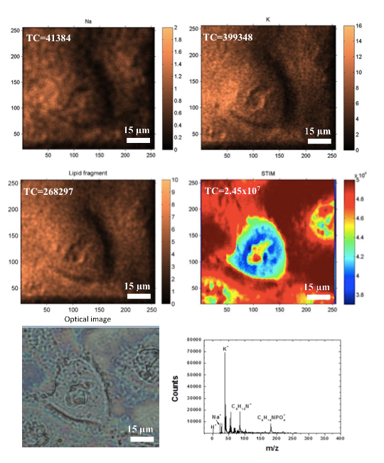
2D distribution of Na, K, and lipids in CaCo-2 cell. A STIM image (density distribution) is also presented, together with an optical image and TOF-SIMS spectrum.
In the case of biomedical applications it is important to perform molecular mapping at the subcellular level. However, the beam spot size of the existing MeV TOF-SIMS systems, which is typically several microns, is too large and has to be reduced. In order to improve the lateral resolution of the focussed beam, the trigger for the TOF – START was replaced with a timing signal provided by a silicon charged particle detector used for Scanning Transmission Ion Microscopy (STIM). By using a trigger signal from detector instead of the bunching the ion microbeam, significant reduction of the object and collimator slit openings was enabled, leading to the reduction of the ion beam spot size (down to 300 nm). Altogether, due to the well-defined submicron beam focus and the high sensitivity, molecular imaging of a single cell at a sub-cellular level has been for the first time achieved by MeV TOF-SIMS as well. Results of this work has been recently published in Applied Physics Letters: Z. Siketić, I. Bogdanović Radović, M. Jakšić, M. Popović Hadžija, M. Hadžija, Submicron mass spectrometry imaging of single cells by combined use of mega electron volt time-of-flight secondary ion mass spectrometry and scanning transmission ion microscopy. In addition, the leading author of the APL paper Zdravko Siketić, presented this work by invited talk at the 13th International Symposium on Radiation Physics held in September in Beijing, China.